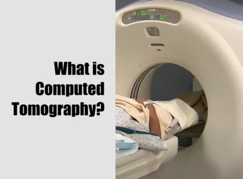
What is Computed Tomography? Computed tomography (CT) is a sophisticated diagnostic imaging technique that uses a combination of X-rays and computer technology to generate detailed cross-sectional images, or slices, of the body’s internal structures. This non-invasive and painless procedure is instrumental in helping healthcare providers diagnose various diseases, injuries, and abnormalities with greater precision and accuracy than conventional X-rays.
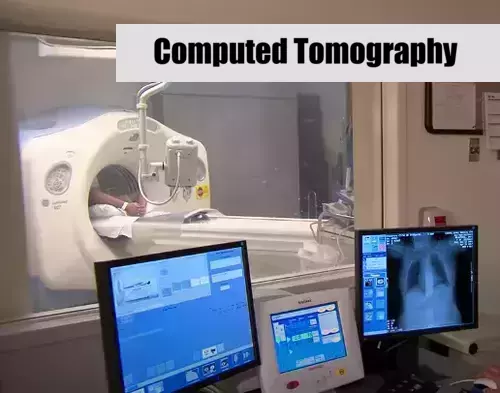
During a CT scan, the patient is positioned on a motorized bed that slowly moves through a doughnut-shaped system called a gantry. The X-ray source within the gantry rotates around the patient, capturing multiple X-ray images from various angles. These images are then sent to a powerful computer that employs advanced mathematical algorithms to construct a series of digital 2D image slices, each representing a specific thickness of the patient’s body, typically ranging from 1 to 10 millimeters.
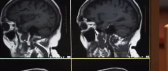
Once the desired number of slices is obtained, they can be digitally combined to create a 3D image of the patient’s body. This comprehensive visual representation allows healthcare providers to examine the patient’s bones, organs, and soft tissues in great detail, identifying potential diseases, injuries, or abnormalities with ease. CT scans have diverse applications, such as detecting tumors, diagnosing heart conditions, examining complex bone fractures, and assessing lung conditions like emphysema, pneumonia, or pulmonary embolisms.
Health Support: This Vitamin K2 + D3 Complex is essential for bone density, cardiovascular health, and immune function. It’s a highly-rated formula for those looking to maintain optimal nutrient levels. You can find it on Amazon.
In some cases, a contrast agent (sometimes referred to as a dye) may be used to enhance the visibility of certain structures during the CT scan, making it easier for healthcare professionals to assess the problem area. The contrast agent, which contains substances that can stop X-rays, is either consumed by the patient or administered intravenously and is eventually eliminated from the body through the urine.
How does CT work?
To understand how CT scans work, let’s start with the scanning process. During a CT scan, the patient lies on a motorized bed that slowly moves through a donut-shaped structure called the gantry. The rotating X-ray source within the gantry shoots narrow beams of X-rays through the patient’s body, while digital detectors opposite the X-ray source pick up the signals and send them to the computer.
The computer uses sophisticated algorithms to turn the X-ray data into two-dimensional image slices, with a thickness typically ranging from 1-10 millimeters. Once the desired number of image slices is collected, the computer can stack them together to form a three-dimensional (3D) image that provides a clear view of the body section being examined. This 3D image can be rotated and viewed from different angles, making it easier to identify abnormalities.
In some cases, contrast agents are used to improve the visibility of internal organs and tissues during a CT scan. These substances, taken orally or injected intravenously, enhance image contrast by stopping X-rays, making certain structures more distinguishable.
Health Support: This high-absorption Magnesium Glycinate (200 mg) is gentle on the stomach and supports muscle relaxation, better sleep, and metabolic health. You can find this trusted formula on Amazon.
Differences between CT and traditional X-ray
One of the key differences between computed tomography (CT) scans and traditional X-ray examinations lies in the level of detail they provide. While both methods use X-rays to create images of the body’s internal structures, CT scans produce cross-sectional images or slices that can be digitally stacked together to create a three-dimensional (3D) image. This enables clinicians to study various structures within the body in much greater detail compared to conventional X-rays, allowing for easier identification of issues such as tumors or abnormalities.
Additionally, the imaging process in a CT scan is more sophisticated. Unlike traditional X-ray machines that use a fixed source, CT scanners utilize a motorized X-ray source that rotates around the patient in a gantry, enabling a more comprehensive view of the targeted area. Furthermore, CT scans use special digital detectors instead of film for improved image quality and easier access to the data.
Another major difference is the utilization of contrast agents in CT scans, providing better visualization of soft tissues that might otherwise be difficult to see with conventional X-rays. Overall, CT scans have advanced imaging capabilities and are particularly useful for detecting issues in complex areas such as bone fractures, eroded joints, and bone tumors where conventional X-rays may not provide sufficient information.
CT contrast agents: what to expect
CT contrast agents are substances used to improve the quality of computed tomography (CT) scan images. These agents, often referred to as “dyes,” help to highlight specific areas or structures within the body, allowing for a more detailed and accurate evaluation by medical professionals. They can be administered orally, via an intravenous injection, or both, depending on the type of CT scan being performed and the reason for the scan.
When preparing for a CT scan with a contrast agent, patients may need to undergo a blood test to determine the appropriate type of dye to be used. It is crucial to follow specific guidelines provided by healthcare providers for food and drink consumption in the hours leading up to the scan. Generally, consuming only clear liquids is recommended to avoid any complications when the contrast agent is administered.
It is important to inform healthcare providers of any allergies to iodine or contrast agents, as this could require medication to be taken before the CT scan to avoid potential allergic reactions. The contrast agent is eliminated from the body through urine within 24 hours after the scan.
How to prepare for a CT scan
Preparing for a CT scan requires following specific guidelines provided by your healthcare provider. Before your appointment, adhere to the following tips:
- Arrive early at the testing facility to avoid delays and ensure a smooth process.
- Refrain from eating and drinking for at least four hours before the examination.
- Consult with your doctor regarding any regular medications you may need to take before the procedure.
- Dress comfortably and be prepared to change into a medical gown if necessary. Remove jewelry, piercings, watches, dentures, and hearing aids as they may affect the results of the scan. Avoid clothing with zippers and metal fastenings.
- If your CT scan uses contrast dye, adhere to any specific preparation guidelines provided by your healthcare provider. This may include a pre-scan blood test, consuming only clear liquids, or taking certain medications if you have a known allergy to the contrast agent.
By adhering to these recommendations, you can help ensure an efficient and accurate CT scan process.
Risks associated with CT scans
One of the main concerns is the exposure to ionizing radiation. Although the amount used during a CT scan is considered minimal, there is a low risk involved with radiation exposure. Pregnant individuals should notify their physicians, as radiation exposure during pregnancy can lead to birth defects.
Another risk comes with the use of contrast dye, which can cause an allergic reaction. If a patient is allergic or sensitive to medications, iodine, or shellfish, they should inform their doctor. For individuals with kidney problems, especially those taking certain diabetic medications, the contrast dye has a potential to cause kidney failure.
Various factors within the body can potentially interfere with the accuracy of a CT scan. Metallic objects in the chest or barium residue from a recent barium study can impact the results. Despite these risks, CT scans remain an essential diagnostic tool when properly conducted and taking patient history into account.
Situations where CT scans may be necessary
CT scans utilize X-ray technology to produce detailed images of the body’s internal structures, including bones, muscles, organs, and blood vessels. By offering a more comprehensive view than standard X-rays, CT scans can significantly enhance a clinician’s ability to diagnose and treat various conditions.
One common situation where a CT scan may be necessary is the assessment of tumor presence, size, and location. This diagnostic method helps determine the need for surgical intervention or other treatments for cancer patients. CT scans are also useful in diagnosing internal bleeding or injuries due to trauma, as they can detect ruptures or damage to organs and blood vessels more effectively than traditional imaging techniques.
Additionally, CT scans play a crucial role in the evaluation of bone fractures and degenerative joint conditions. They can provide intricate details of the affected area, helping healthcare professionals develop appropriate treatment plans. In cases of suspected heart disease or abnormalities, a CT scan can be invaluable in precisely pinpointing the problem and guiding subsequent interventions.
The use of CT scans in the detection of lung-related conditions, such as pulmonary embolisms, excess fluid, and pneumonia, is another crucial application. Their ability to offer clear and detailed images assists in the successful diagnosis and treatment of these potentially serious issues. By understanding the various situations where CT scans may be necessary, patients and healthcare professionals alike can better utilize this advanced diagnostic tool to ensure optimal health outcomes.
What diseases can be detected by CT scanning
Some of the diseases that can be detected by CT scanning are:
- Cancer: CT scans can detect various types of cancerous tumors, including lung, brain, liver, and pancreatic cancer, by revealing the size, shape, and location of the abnormal growths.
- Internal injuries and bleeding: CT scans can be used to identify internal injuries from traumatic events such as car accidents, falls, or sports injuries. These scans help detect internal bleeding, organ damage, and fractures that may not have surfaced yet.
- Infections and inflammatory diseases: CT scans can detect conditions such as abscesses, sinusitis, and pneumonia by revealing areas of inflammation and infection in the body.
- Vascular diseases: CT scans can visualize blood vessels and detect blockages, narrowing, or aneurysms that may lead to severe conditions like strokes or heart attacks.
- Kidney and bladder stones: CT scans efficiently detect kidney and bladder stones, their size, shape, and location, aiding in deciding the best treatment.
- Skeletal disorders: The imaging provided by CT scans can diagnose issues like bone tumors, complex fractures, or arthritis, offering detailed information about the structure and possible damage to the bones.
- Lung diseases: CT scans are useful for detecting lung diseases like chronic obstructive pulmonary disease (COPD), emphysema, and tuberculosis by providing clear images of the lungs and associated abnormalities.
Advantages and limitations of CT
Computed tomography (CT) scans are a valuable diagnostic tool in the medical field, but they also have some limitations and risks.
One of the key advantages of CT scans is their ability to produce detailed cross-sectional images of internal organs, bones, soft tissues, and blood vessels. These images provide greater clarity and detail than conventional x-ray images, enabling physicians to plan and guide interventional or therapeutic procedures, monitor the effectiveness of therapy and diagnose a wide range of conditions in adults and children.
In many cases, the detailed information provided by CT scans can eliminate the need for exploratory surgery. CT scans are noninvasive and are usually performed as outpatient procedures, which means that patients do not need to be hospitalized for the examination.
However, there are also some drawbacks to CT scans. One of the major concerns is the exposure to ionizing radiation, which may cause a small increase in a person’s lifetime risk of developing cancer. The risk is particularly higher for young patients, as their bodies are more sensitive to radiation and they have a longer lifetime for the effects of radiation exposure to manifest as cancer.
Another limitation is the cost of CT examinations, which can be quite expensive and may create a significant financial burden for patients, especially those without medical insurance or those who undergo multiple scans.
Lastly, the misuse and overreliance on CT scans can promote laziness in medical professionals, as these tests might sometimes be ordered indiscriminately without considering alternative diagnostic methods. This could lead to misdiagnosis or misinterpretation of CT scan images.
CT vs MRI: what’s the difference?
Computed tomography (CT) and magnetic resonance imaging (MRI) are two distinct yet complementary medical imaging techniques utilized by doctors to diagnose various health conditions. These non-invasive methods provide valuable insights about the internal structures and abnormalities in patients without the need for surgery or invasive procedures.
A CT scan uses a series of X-rays taken in rapid succession from different angles to create a detailed, three-dimensional image of a patient’s body. On the other hand, MRI employs strong magnets and radio waves to generate pictures of body structures. While CT scans excel in defining the edges and boundaries of structures within the body, MRIs provide superior differentiation between various tissue types, making it easier to distinguish between healthy and cancerous tissues.
When deciding which imaging method to use, doctors consider the patient’s specific health condition, the type and location of the suspected problem, and the overall objective of the examination. CT scans might be a better choice when assessing bony structures, while MRIs are more suitable for mapping soft tissues, such as ligaments, joints, or internal organs.
One of the significant differences between CT and MRI scans is their duration: CT scans are completed within minutes, whereas MRI scans generally take 30 minutes to an hour. This difference in duration can be a factor for patients who have difficulty staying still for long periods or those experiencing severe pain. However, both techniques are essential diagnostic tools that provide clinicians with valuable information to accurately diagnose and treat patients.
Average price for a CT scan
| Clinic Name | Average Price for CT Scan in 2023 (in USD) |
|---|---|
| Mayo Clinic | 1,200 |
| Cleveland Clinic | 1,350 |
| Johns Hopkins Hospital | 1,500 |
| Mass General Hospital | 1,600 |
| UCLA Medical Center | 1,700 |
| Mount Sinai Hospital | 1,800 |
| NYU Langone Hospital | 1,900 |
| Cedars-Sinai Medical Center | 2,000 |
The average national price for a CT scan ranges from $300 to $6,750. Some health plans may partially or fully cover authorized CT scans, but patients may still have out-of-pocket costs such as copays, coinsurance, or deductibles. Costs can differ between inpatient facilities (hospitals) and outpatient centers, with the latter often being more affordable. In-network providers or facilities that have a contract with a health insurance company typically offer lower rates compared to out-of-network providers.
It is crucial for individuals to consult with their healthcare provider and insurance company to better understand any related expenses before undergoing a CT scan. For patients without insurance, researching and comparing prices at various facilities may help in finding an affordable option, ultimately ensuring access to this vital diagnostic imaging tool.