Histology and cytology can make a lot of sense when it comes to someone’s medical condition. These terms are not always clear to people who are not familiar with medicine. A reasonable question arises: what is the difference between histology and cytology? Let’s try to get to the bottom of it.
Definition
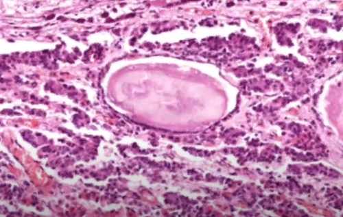
Histology is a discipline dedicated to the study of tissues of various organisms, including human beings. This is also the name of the very process of studying the named biological material.
Cytology is the science of what forms the basis of the structure of all living things, that is, cells. The same word means a method that involves the study of such structural units within the laboratory.
Comparison
Each case has its own object of consideration, which is the main difference between histology and cytology. Thus, the first of these concepts has to do with tissues, their structure and functions performed. Cytology, on the other hand, is focused on the study of structures on a smaller scale – cellular.
To perform a histological study, it is first necessary to remove a fragment of the desired tissue from the body. This may require a biopsy. Sometimes it is taken simultaneously with surgical intervention. The obtained biological material is prepared in several stages, and then the obtained preparation is carefully studied under a microscope. The results will be the basis for making a diagnosis.
What is the difference between histology and cytology? The first method is invasive and is usually applied when the disease has already made itself felt. Meanwhile, cytology is performed without any trauma to the body. However, this procedure allows you to recognize a pathology that is only incipient, even in the absence of alarming symptoms. First of all it concerns such a disease as cancer.
The simplest cytological examination involves taking a smear, placing it on a glass and drying it, after which the material is stained and examined under high magnification. Conclusions about the development of the disease in this case are made on the basis of observed changes in the cellular structure.
The two examinations considered are often carried out one after another: first, the tissue as a whole is examined, and then a deeper, cellular analysis of the material is performed. Sometimes there is no need for histological intervention and only cytology can be done. For example, to find out if a uterine erosion has developed, it is enough to examine a smear.
About the Author
Reyus Mammadli is the author of this health blog since 2008. With a background in medical and biotechnical devices, he has over 15 years of experience working with medical literature and expert guidelines from WHO, CDC, Mayo Clinic, and others. His goal is to present clear, accurate health information for everyday readers — not as a substitute for medical advice.


