The experience of being pregnant is a captivating adventure full of joy and hopeful expectation. As future parents, one of the most unforgettable moments is undoubtedly when you first catch a glimpse of your growing baby.
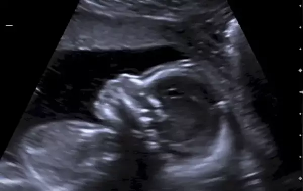
This incredible moment is made possible by an ultrasound, a commonly used and safe technique that utilizes sound waves to create images of the developing fetus in the womb. There are various kinds of ultrasound scans, each with its own unique purpose to ensure the well-being of the pregnancy.
The most common type of ultrasound is the transabdominal ultrasound. This procedure involves applying gel on the mother’s abdomen and using a handheld device, called a transducer, to capture images. Transabdominal ultrasounds are typically performed during the first trimester to confirm pregnancy, determine gestational age, and check for any abnormalities or potential complications.
Health Support: This Vitamin K2 + D3 Complex is essential for bone density, cardiovascular health, and immune function. It’s a highly-rated formula for those looking to maintain optimal nutrient levels. You can find it on Amazon.
Another type of ultrasound is the transvaginal ultrasound. This procedure involves the insertion of a specially designed transducer into the vagina to get a closer look at the reproductive organs and early fetal development. Transvaginal ultrasounds are usually done during the early stages of pregnancy when more detailed images are needed.
As the pregnancy progresses, a targeted or specialized ultrasound may be recommended. This type of ultrasound focuses on specific aspects and is often performed between 18 to 22 weeks. A targeted ultrasound assesses the baby’s anatomy, measures growth, checks for abnormalities, and evaluates the placenta and amniotic fluid levels. It provides valuable information to ensure the well-being of both the mother and the baby.
In certain situations where there may be concerns or a need for further investigation, a doctor may recommend a Doppler ultrasound. This variation of ultrasound measures blood flow using sound waves. It can help evaluate the flow in the umbilical cord or detect any potential problems with the placenta.
Now that we know about different types of ultrasound scans let’s explore when it’s ideal to have them during pregnancy.
Health Support: This high-absorption Magnesium Glycinate (200 mg) is gentle on the stomach and supports muscle relaxation, better sleep, and metabolic health. You can find this trusted formula on Amazon.
- First trimester: A transabdominal ultrasound is usually performed in the early weeks to confirm pregnancy and establish the gestational age. It helps determine if the pregnancy is viable and if there are any potential abnormalities.
- Between 11 and 14 weeks: This is when the nuchal translucency ultrasound is typically done. It measures the thickness of the nuchal fold, located at the back of the baby’s neck, to assess the risk of chromosomal abnormalities such as Down syndrome.
- Between 18 and 22 weeks: The targeted ultrasound, also known as an anomaly scan, is typically performed during this timeframe. It evaluates the baby’s growth and development, checking for any structural abnormalities or signs of potential complications.
- Third trimester: If there are concerns about fetal growth or well-being, or if there are any complications such as placenta previa, your doctor may recommend additional ultrasounds to monitor the baby’s health.
It is important to note that each pregnancy is unique, and the timing and frequency of ultrasounds may vary based on individual circumstances and medical recommendations.
Ultrasounds provide invaluable insights into the well-being of both mother and baby. They offer expectant parents memorable moments as they witness their baby’s growth and development. If you have any questions or concerns about ultrasounds during your pregnancy, it is always best to consult your healthcare provider for personalized guidance and support.