What is Relapsing Perichondritis?
Relapsing polychondritis is an unusual degenerative disease characterized by frequent inflammation of the cartilage in the body.
Wear and tear of the cartilage may impact any site of the body where cartilage exists. Ears, larynx and trachea might become “floppy,” and the bridge of the nose can collapse into a “saddlenose” shape. The aortic heart valve may also be affected.
More Details about Relapsing Perichondritis
- Common cartilage tissues affected consist of the ears, nose, and joints.
- Relapsing polychondritis is an unusual, chronic disorder of the cartilage.
- Relapsing polychondritis is defined by reoccurring episodes of painful inflammation.
- Treatment frequently involves cortisone-related medications.
- Relapsing polychondritis can involve all types of cartilage.
- There is nobody particular test for diagnosing relapsing polychondritis.
- The course of symptoms for patients is frequently unpredictable.
Symptoms of Relapsing Perichondritis
Injuries, burns, insect bites, ear piercings through the cartilage, ear surgery, or a boil on the ear might trigger perichondritis. The infection also tends to happen in people who have inflammatory disorders, such as granulomatosis with polyangiitis, whose immune system is weakened, or who have diabetes.
Health Support: This Vitamin K2 + D3 Complex is essential for bone density, cardiovascular health, and immune function. It’s a highly-rated formula for those looking to maintain optimal nutrient levels. You can find it on Amazon.
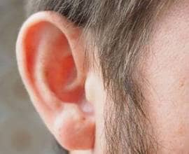
The symptoms of Relapsing Perichondritis same as Perichondritis: usually starts from inflammation, pain, and swelling of the auricle. The person may have a fever. Pus accumulates in between the cartilage and the layer of connective tissue around it (perichondrium). Sometimes the pus cuts off the blood supply to the cartilage, ruining it and leading eventually to a warped ear (also known as cauliflower ear). Perichondritis can be destructive, repeat, and last for weeks and months.
Causes
The specific reason for relapsing polychondritis is not known. It is believed to be an autoimmune disease. Autoimmune conditions are triggered when the body’s natural defenses against “foreign” or invading organisms (e.g., antibodies) start to assault healthy tissue for unknown reasons. Some cases might be linked to irregular reactions by blood cells (serum antibodies), to a thyroid protein (thyroglobulin), organ wall (parietal) cells, adrenal cells, or thyroid. Symptoms of relapsing polychondritis may develop when autoantibodies attack human cartilage.
Some scientists think that relapsing polychondritis may be triggered by an immunologic level of sensitivity to type II collagen, a normal substance found in skin and connective tissue.
Affected Populations
Relapsing polychondritis affects males and females in equivalent numbers. Symptoms usually start in between forty and sixty years of age.
Health Support: This high-absorption Magnesium Glycinate (200 mg) is gentle on the stomach and supports muscle relaxation, better sleep, and metabolic health. You can find this trusted formula on Amazon.
Associated Disorders
Symptoms of the following disorders can be similar to those of relapsing polychondritis. Contrasts might work for a differential diagnosis:
Rheumatoid arthritis is a disease of unknown origin which might have a relationship to autoimmune procedures. This condition is defined by lack of cravings (anorexia), exhaustion, painful and warped joints, early morning stiffness chiefly in the hands, knees, feet, jaw, and spine. As soon as affected, a patient’s joints stay painful or unpleasant for weeks, months, and even years.
Osteoarthritis is a degenerative joint disease of unidentified origin characterized by loss of cartilage, defects of bones with joints, and additional cartilage and bone growth at the joint margins with subsequent bony enlargement. Osteoarthritis develops when cartilage repair does not equal degeneration. It might take place as a result of injury to the bony or underlying joint disease.
Behcet syndrome is an inflammatory disorder impacting a variety of organs. The most consistent sign is of oral and genital ulcers. Eye and joint inflammation, similar to Polychondritis, happens. Blood vessels, the central nerve system, and the gastrointestinal tract might likewise be involved. Attacks last a week to a month, and can repeat spontaneously. Some symptoms can appear as late as a number of years after beginning of the disease which usually takes place in between age 20 and 30. Two times as lots of men as women are impacted. The disease is most typical in the Middle East and Japan. For additional information on the above disorder, select “Behcet” as your search term in the Rare Disease Database.
How Does Relapsing Perichondritis Treated
Treatment of relapsing polychondritis generally includes the administration of corticosteroid drugs (e.g., prednisone), aspirin and non-steroidal anti-inflammatory compounds such as dapsone and/or colchicine. In extreme cases, drugs that reduce the immune system such as cyclophosphamide, 6-mercaptopurine and azathioprine might be advised. In the most extreme cases replacement of heart valves or the insertion of a breathing tube (tracheotomy) for collapsed airways might be essential.
- Antibiotics or, maybe, corticosteroids
- Removal of foreign items, particularly ear piercings through the cartilage part of the auricle
- Warm compresses and incision and drain of abscesses
- Painkiller
Medical professionals deal with perichondritis with antibiotics (such as a fluoroquinolone, for instance, ciprofloxacin) and typically a corticosteroid by mouth. The option of antibiotic depends on how serious the infection is and which bacteria are triggering it.
Medical professionals remove any foreign objects, such as an earring or a splinter.
If individuals have an abscess (collection of pus), medical professionals make an incision to drain the pus, enabling blood to reach the cartilage again, and leave a small drain in place for 24 to 72 hours. Antibiotics are given by mouth. Warm compresses may also assist. Doctors may sew (stitch) the perichondrium to the cartilage to make sure that it recovers properly to prevent a deformity of the auricle.
Pain relievers are also offered.
Story of Patient with Perichondritis
A 30-year-old woman provided with grievances of a swollen right pinna for 10 days. The swelling progressively gotten worse in time. In addition, she also experienced extreme pain of the right pinna, with an intensity of 7/10, without any radiation and no aggravating or alleviating elements. One week previously, she had actually been taken a look at in an urgent care facility and was offered a 10-day course of amoxicillin for presumed acute otitis externa. However, the infection continued to intensify. There was no history of injury to the ear and no other substantial case history.
On examination, the woman had an irritated, erythematous and tender right pinna. The pr- and post-auricular lymph nodes were enlarged and tender. Evaluation of the remainder of the ear was normal and hearing was not impaired. She was afebrile and all other systemic evaluations were normal.