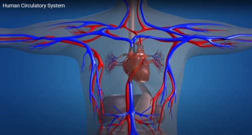
In the circulatory system, there are several types of blood vessels that link the venous system to the arterial system. Arteries are the pathways through which blood travels away from the heart, while veins transport blood back to the heart.
Arteries and veins have three layers:
- The external layer consists of connective tissue.
- The middle section of the vessel, made up of muscle that is smooth and elastic fibers, encircles the inner space of the vessel.
- The inner layer is made up of collagen and elastic fibers as well as the endothelium.
Arteries
Due to the high-pressure flow of blood from the heart to the arteries, the arteries possess thicker walls and greater elasticity compared to veins. The aorta, the largest artery, transports blood away from the heart. As it moves away from the heart, the aorta divides into progressively smaller vessels that deliver blood to all organs and tissues, eventually forming a network of capillaries.
Arterial blood is a vibrant red shade and contains substantial quantities of oxygen carried by red blood cell hemoglobin. Additionally, it contains energy-rich substances vital for cellular function, with only a small quantity of oxygen dissolved in the blood.
Veins
The journey of the venous system starts in the venules, where blood collects waste products from nearby tissues. It then moves through increasingly wider veins to reach the right atrium of the heart.
The heart receives venous blood from the larger circulatory system through two major vessels: the inferior vena cava and the superior vena cava.
Because the pressure of the blood in the veins is low, their walls do not require the same thickness and elasticity as the arteries.
Moreover, the veins have valves within their inner space to prevent backward flow of blood. Venous blood, which lacks oxygen, appears darker in color and carries waste products and carbon dioxide (largely dissolved within the blood).
Capillary system
The structure is made up of a close-knit network of small vessels located between the arterial and venous systems that weave through all tissues.
These vessels have walls made up of only one layer of cells. This particular structure enables close proximity of blood to cells, enables gas exchange, facilitates the delivery of nutrients to cells, and aids in the removal of waste products from cells.
Comparison table of vein, artery, and capillary functions
| Veins | Arteries | Capillaries |
|---|---|---|
| Carry deoxygenated blood towards the heart. | Carry oxygenated blood away from the heart. | Allow the exchange of nutrients, gases, and waste products between the blood and surrounding tissues. |
| Have thin walls and valves to prevent backflow of blood. | Have thick walls with elastic fibers to withstand high pressure. | Have the thinnest walls, consisting of a single layer of cells, facilitating the diffusion of substances. |
| Have larger lumen and can expand to hold a larger volume of blood. | Have smaller lumen and maintain a high pressure to propel blood forward. | Have narrow and numerous capillary beds, increasing the surface area for efficient exchange. |
| Are less muscular compared to arteries. | Have thick smooth muscle layers, allowing them to constrict and dilate to regulate blood flow. | Are mainly responsible for the exchange of substances between blood and tissues. |
| Aid in the return of blood back to the heart, aided by valves and skeletal muscle contractions. | Distribute blood to various organs and tissues throughout the body. | Connect arteries and veins, forming a network within tissues for exchange. |
| Have lower blood pressure compared to arteries. | Have higher blood pressure to maintain efficient blood flow. | Play a crucial role in maintaining homeostasis and supplying tissues with oxygen and nutrients. |
| Examples include the superior vena cava and pulmonary veins. | Examples include the aorta and coronary arteries. | Present in various organs such as the lungs, liver, and kidneys. |
About the Author
Reyus Mammadli is the author of this health blog since 2008. With a background in medical and biotechnical devices, he has over 15 years of experience working with medical literature and expert guidelines from WHO, CDC, Mayo Clinic, and others. His goal is to present clear, accurate health information for everyday readers — not as a substitute for medical advice.


